|
|
Metastases to Bone
Renal Cell Carcinoma
General Considerations
- Metastases are most common malignant bone tumors
- Most involve axial skeleton
- Skull, spine and pelvis
- Rarely do mets occur distal to elbows or knees
- Spread hematogenously
- Most frequently occur where red bone marrow is found
- Mets to spine frequently destroy posterior vertebral body including
pedicle first=”pedicle-sign”
- 90% of skeletal mets are multiple
- Primary carcinomas that frequently metastasize to bone
- The next four lesions comprise 80% of all metastases to bone
- Breast (70% of bone mets in women)
- Lung
- Prostate (60% of all bone mets in men)
- Kidney
- Also
- Thyroid
- Stomach and intestines
- Clinical
- Most lesions are asymptomatic
- When symptomatic, pain is major symptom
- Fractures of the lesser trochanter in adults should be considered
pathologic until proven otherwise
- Imaging Findings
- In general, mets have little or no soft tissue mass associated with them
- Usually no periosteal reaction
- May appear as moth-eaten, permeative or geographic lesions
- Indistinct zones of transition
- No sclerotic margins
- May be expansile
- Soap-bubbly (septated)
- May be sharply circumscribed or have indistinct borders
- Metastases that are typically purely lytic
- Metastases that are usually mixed lytic and sclerotic
- Metastases that are usually purely blastic
- Prostate
- Medulloblastoma
- Bronchial carcinoid
- No matter what the primary, skull metastases are usually lytic in appearance
Most Common Tumors to Metastasize to Bone(80% of bone mets) |
| Prostate |
Blastic |
| Breast |
Mixed |
| Lung |
Predominantly lytic |
| Renal Cell Ca |
Predominantly Lytic |
- Imaging findings suggestive of a particular primary tumor
- Lesions distal to elbows and knees
- 50% are from lung and breast
- Expansile and lytic (soap-bubbly)
- Diffuse skeletal sclerosis or multiple round, well-circumscribed
sclerotic lesions
- Cookie-bite lesions of the cortices of long bones
- Radioscintographic studies
- Bone scans are extremely sensitive but not very specific
- 10-40% of lesions will not be visible on plain film but will be positive
on bone scans
- CT or MRI can be used to show findings in patients with negative
conventional radiographs and positive bone scans
- Complications of metastases to bone
- Pathologic fractures
- Destruction of 50% or more of bone suggests impending
pathologic fracture
- Spinal cord compression
- Treated lytic mets may become sclerotic with treatment

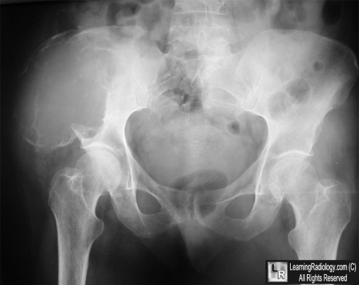
Metastases from Renal Cell Carcinoma. Frontal radiograph of the pelvis demonstrates a
huge, expansile osteolytic lesion destroying most of right ilium (yellow arrows). The lesion contains septa. Expansile metastases such as this generally occur with renal cell and thyroid carcinomas.
For this same photo without arrows, click here
For more information, click on the link if you see this icon 
Orthopedic radiology: A Practical Approach, Greenspan, Ada; Lippincott, 2000
Diagnosis of Bone and Joint Disorders, Resnick, Donald, W. B. Saunders
Musculoskeletal Imaging: The Requisites, Manaster, BJ et al; Mosby, 2002
|
|
|
